

Fungi And Animals Are Both Found In Which Eukaryotic Supergroup?
Protists
118 Groups of Protists
Learning Objectives
Past the stop of this section, you will be able to exercise the following:
- Describe representative protist organisms from each of the six presently recognized supergroups of eukaryotes
- Identify the evolutionary relationships of plants, animals, and fungi inside the half-dozen soon recognized supergroups of eukaryotes
- Identify defining features of protists in each of the half dozen supergroups of eukaryotes.
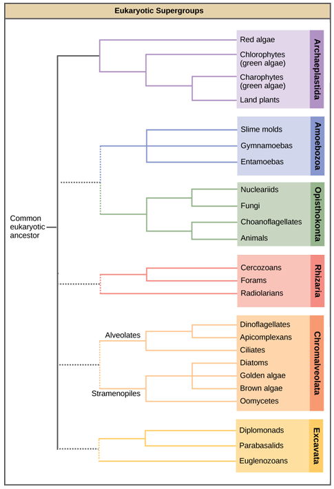
In the span of several decades, the Kingdom Protista has been disassembled because sequence analyses have revealed new genetic (and therefore evolutionary) relationships among these eukaryotes. Moreover, protists that exhibit like morphological features may accept evolved analogous structures because of similar selective pressures—rather than because of recent common beginnings. This phenomenon, called convergent evolution, is one reason why protist classification is and then challenging. The emerging classification scheme groups the entire domain Eukarya into 6 "supergroups" that contain all of the protists also as animals, plants, and fungi that evolved from a common ancestor ((Figure)). Each of the supergroups is believed to be monophyletic, meaning that all organisms within each supergroup are believed to accept evolved from a single common ancestor, and thus all members are most closely related to each other than to organisms exterior that grouping. In that location is notwithstanding evidence lacking for the monophyly of some groups. Each supergroup can be viewed every bit representing 1 of many variants on eukaryotic cell structure. In each grouping, ane or more than of the defining characters of the eukaryotic cell—the nucleus, the cytoskeleton, and the endosymbiotic organelles—may take diverged from the "typical" blueprint.
Eukaryotic supergroups. This diagram shows a proposed classification of the domain Eukarya. Currently, the domain Eukarya is divided into half dozen supergroups. Inside each supergroup are multiple kingdoms. Although each supergroup is believed to be monophyletic, the dotted lines suggest evolutionary relationships among the supergroups that go on to exist debated.
Keep in mind that the classification scheme presented here represents simply one of several hypotheses, and the true evolutionary relationships are still to exist determined. The six supergroups may be modified or replaced by a more appropriate hierarchy as genetic, morphological, and ecological data accumulate. When learning virtually protists, it is helpful to focus less on the classification and more than on the commonalities and differences that illustrate how each group has exploited the possibilities of eukaryotic life.
Archaeplastida
Molecular evidence supports the hypothesis that all Archaeplastida are descendents of an endosymbiotic relationship betwixt a heterotrophic protist and a cyanobacterium. The protist members of the group include the red algae and green algae. It was from a common ancestor of these protists that the land plants evolved, since their closest relatives are found in this group. The red and green algae include unicellular, multicellular, and colonial forms. A variety of algal life cycles exists, but the about complex is alternation of generations, in which both haploid and diploid stages are multicellular. A diploid sporophyte contains cells that undergo meiosis to produce haploid spores. The spores germinate and grow into a haploid gametophyte, which then makes gametes by mitosis. The gametes fuse to course a zygote that grows into a diploid sporophyte. Alternation of generations is seen in some species of Archaeplastid algae, as well as some species of Stramenopiles ((Effigy)). In some species, the gametophyte and sporophyte look quite different, while in others they are near duplicate.
Glaucophytes
Glaucophytes are a small group of Archaeplastida interesting considering their chloroplasts retain remnants of the peptidoglycan jail cell wall of the ancestral cyanobacterial endosymbiont ((Effigy)).
Red Algae
Carmine algae, or rhodophytes lack flagella, and are primarily multicellular, although they range in size from microscopic, unicellular protists to large, multicellular forms grouped into the informal seaweed category. Red algae have a second cell wall outside an inner cellulose cell wall. Carbohydrates in this wall are the source of agarose used for electrophoresis gels and agar for solidifying bacterial media. The "red" in the scarlet algae comes from phycoerythrins, accessory photopigments that are ruby-red in colour and obscure the greenish tint of chlorophyll in some species. Other protists classified every bit red algae lack phycoerythrins and are parasites. Both the red algae and the glaucophytes shop carbohydrates in the cytoplasm rather than in the plastid. Ruby-red algae are common in tropical waters where they have been detected at depths of 260 meters. Other red algae exist in terrestrial or freshwater environments. The cherry algae life cycle is an unusual alternation of generations that includes two sporophyte phases, with meiosis occurring only in the second sporophyte.
Green Algae: Chlorophytes and Charophytes
The most arable group of algae is the light-green algae. The green algae showroom features similar to those of the land plants, especially in terms of chloroplast structure. In both green algae and plants, carbohydrates are stored in the plastid. That this group of protists shared a relatively recent mutual antecedent with state plants is well supported. The greenish algae are subdivided into the chlorophytes and the charophytes. The charophytes are the closest living relatives to land plants and resemble them in morphology and reproductive strategies. The familiar Spirogyra is a charophyte. Charophytes are common in moisture habitats, and their presence often signals a healthy ecosystem.
The chlorophytes exhibit neat diversity of class and office. Chlorophytes primarily inhabit freshwater and damp soil, and are a common component of plankton. Chlamydomonas is a simple, unicellular chlorophyte with a pear-shaped morphology and 2 opposing, anterior flagella that guide this protist toward light sensed by its eyespot. More complex chlorophyte species showroom haploid gametes and spores that resemble Chlamydomonas.
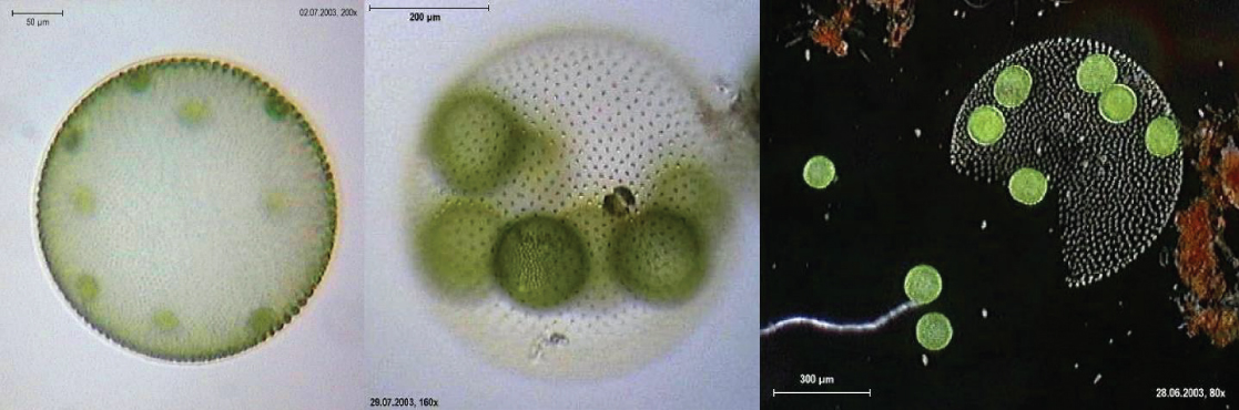
The chlorophyte Volvox is one of only a few examples of a colonial organism, which behaves in some means like a collection of private cells, merely in other ways similar the specialized cells of a multicellular organism ((Effigy)). Volvox colonies contain 500 to 60,000 cells, each with 2 flagella, contained within a hollow, spherical matrix composed of a gelatinous glycoprotein secretion. Individual cells in a Volvox colony move in a coordinated manner and are interconnected by cytoplasmic bridges. Simply a few of the cells reproduce to create girl colonies, an example of bones cell specialization in this organism. Daughter colonies are produced with their flagella on the inside and take to evert as they are released.
Volvox. Volvox aureus is a green alga in the supergroup Archaeplastida. This species exists equally a colony, consisting of cells immersed in a gel-like matrix and intertwined with each other via hair-like cytoplasmic extensions. (credit: Dr. Ralf Wagner)
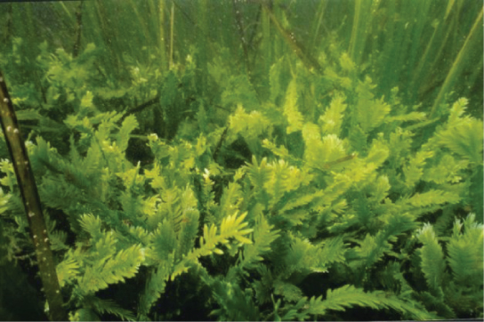
Truthful multicellular organisms, such every bit the body of water lettuce, Ulva, are also represented among the chlorophytes. In addition, some chlorophytes exist as big, multinucleate, single cells. Species in the genus Caulerpa exhibit flattened fern-like leafage and tin can achieve lengths of 3 meters ((Effigy)). Caulerpa species undergo nuclear division, just their cells do not complete cytokinesis, remaining instead equally massive and elaborate unmarried cells.
A multinucleate alga. Caulerpa taxifolia is a chlorophyte consisting of a single cell containing potentially thousands of nuclei. (credit: NOAA). An interesting question is how a single cell tin produce such circuitous shapes.
Link to Learning
Take a look at this video to see cytoplasmic streaming in a light-green alga.
Amoebozoa
Like the Archaeplastida, the Amoebozoa include species with single cells, species with large multinucleated cells, and species that accept multicellular phases. Amoebozoan cells characteristically exhibit pseudopodia that extend like tubes or flat lobes. These pseudopods project outward from anywhere on the cell surface and can anchor to a substrate. The protist and so transports its cytoplasm into the pseudopod, thereby moving the entire prison cell. This blazon of motion is similar to the cytoplasmic streaming used to move organelles in the Archaeplastida, and is as well used past other protists as a means of locomotion or as a method to distribute nutrients and oxygen. The Amoebozoa include both complimentary-living and parasitic species.
Gymnomoebae
The Gymnamoeba or lobose amoebae include both naked amoebae like the familiar Amoeba proteus and shelled amoebae, whose bodies protrude like snails from their protective tests. Amoeba proteus is a large amoeba about 500 µm in diameter simply is dwarfed past the multinucleate amoebae Pelomyxa, which can be ten times its size. Although Pelomyxa may accept hundreds of nuclei, it has lost its mitochondria, merely replaced them with bacterial endosymbionts. The secondary loss or modification of mitochondria is a feature also seen in other protist groups.
Amoeba. Amoebae with tubular and lobe-shaped pseudopodia are seen under a microscope. These isolates would be morphologically classified as amoebozoans.
Slime Molds
A subset of the amoebozoans, the slime molds, has several morphological similarities to fungi that are thought to be the result of convergent development. For instance, during times of stress, some slime molds develop into spore-generating fruiting bodies, much like fungi.
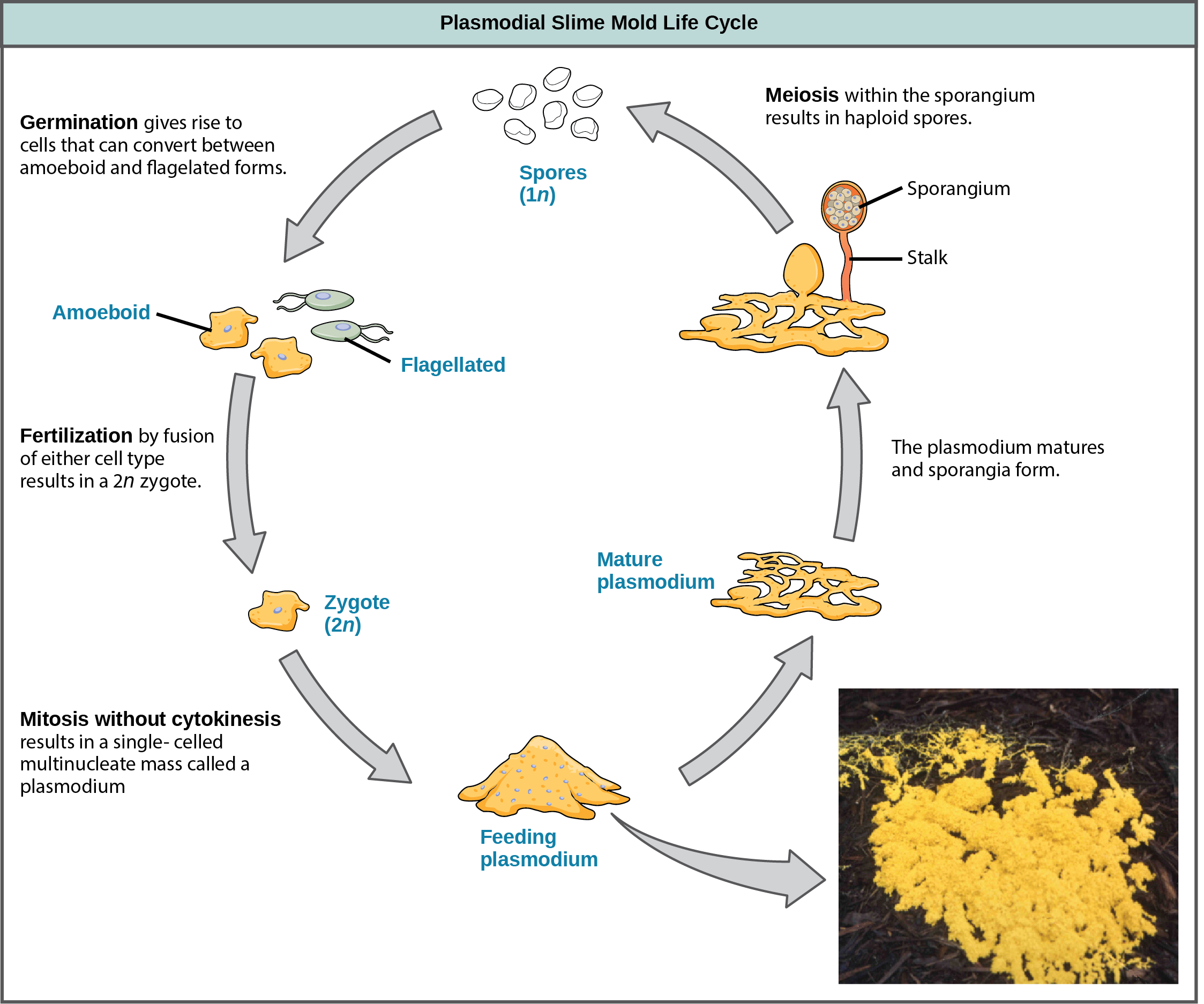
The slime molds are categorized on the basis of their life cycles into plasmodial or cellular types. Plasmodial slime molds are composed of big, multinucleate cells and motion forth surfaces like an amorphous blob of slime during their feeding phase ((Figure)). Food particles are lifted and engulfed into the slime mold as information technology glides along. The "dog vomit" slime mold seen in (Figure) is a particularly colorful specimen and its ability to creep about might well trigger suspicion of alien invasion. Upon maturation, the plasmodium takes on a net-similar advent with the power to form fruiting bodies, or sporangia, during times of stress. Haploid spores are produced by meiosis within the sporangia, and spores can be disseminated through the air or h2o to potentially land in more favorable environments. If this occurs, the spores germinate to form ameboid or flagellate haploid cells that can combine with each other and produce a diploid zygotic slime mold to consummate the life cycle.
Plasmodial slime molds. The life cycle of the plasmodial slime mold is shown. The brightly colored plasmodium in the inset photo is a unmarried-celled, multinucleate mass. (credit: modification of piece of work by Dr. Jonatha Gott and the Center for RNA Molecular Biology, Case Western Reserve Academy)
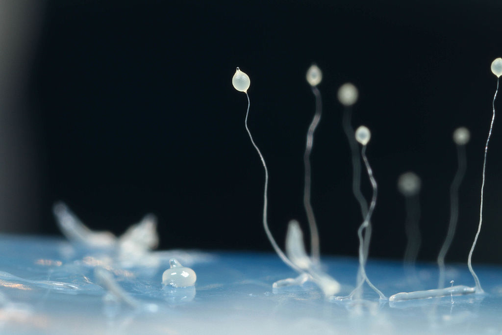
The cellular slime molds function as independent amoeboid cells when nutrients are abundant. When nutrient is depleted, cellular slime molds aggregate into a mass of cells that behaves as a single unit, called a slug. Some cells in the slug contribute to a two–3-millimeter stalk, drying upwards and dying in the process. Cells atop the stalk form an asexual fruiting body that contains haploid spores ((Effigy)). As with plasmodial slime molds, the spores are disseminated and can germinate if they state in a moist environment. One representative genus of the cellular slime molds is Dictyostelium, which commonly exists in the damp soil of forests.
Cellular Slime Mold. The prototype shows several stages in the life cycle of Dictyostelium discoideum, including aggregated cells, mobile slugs and their transformation into fruiting bodies with a cluster of spores supported past a stalk. (credit: By Usman Bashir (Own piece of work) [CC Past-SA 4.0 (http://creativecommons.org/licenses/by-sa/4.0)], via Wikimedia Commons)
Link to Learning
View this video to see the germination of a fruiting torso past a cellular slime mold.
Opisthokonta
The Opisthokonts are named for the single posterior flagellum seen in flagellated cells of the grouping. The flagella of other protists are inductive and their movement pulls the cells along, while the opisthokonts are pushed. Protist members of the opisthokonts include the animal-like choanoflagellates, which are believed to resemble the common ancestor of sponges and perchance, all animals. Choanoflagellates include unicellular and colonial forms ((Figure)), and number most 244 described species. In these organisms, the single, apical flagellum is surrounded by a contractile collar composed of microvilli. The collar is used to filter and collect bacteria for ingestion by the protist. A similar feeding machinery is seen in the collar cells of sponges, which suggests a possible connection between choanoflagellates and animals.
The Mesomycetozoa form a small group of parasites, primarily of fish, and at least one form that tin can parasitize humans. Their life cycles are poorly understood. These organisms are of special interest, because they announced to be and then closely related to animals. In the by, they were grouped with fungi and other protists based on their morphology.
The previous supergroups are all the products of primary endosymbiontic events and their organelles—nucleus, mitochondria, and chloroplasts—are what would exist considered "typical," i.e., matching the diagrams you lot would detect in an introductory biology book. The next three supergroups all contain at least some photosynthetic members whose chloroplasts were derived by secondary endosymbiosis. They besides evidence some interesting variations in nuclear construction, and modification of mitochondria or chloroplasts.
Rhizaria
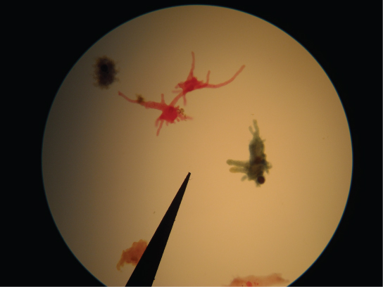
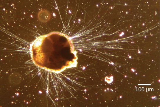
The Rhizaria supergroup includes many of the amoebas with thin threadlike, needle-like or root-like pseudopodia ((Figure)), rather than the broader lobed pseudopodia of the Amoebozoa. Many rhizarians make elaborate and beautiful tests—armor-like coverings for the body of the cell—composed of calcium carbonate, silicon, or strontium salts. Rhizarians take of import roles in both carbon and nitrogen cycles. When rhizarians die, and their tests sink into deep water, the carbonates are out of attain of about decomposers, locking carbon dioxide away from the atmosphere. In full general, this process by which carbon is transported deep into the ocean is described as the biological carbon pump, considering carbon is "pumped" to the ocean depths where information technology is inaccessible to the temper as carbon dioxide. The biological carbon pump is a crucial component of the carbon cycle that maintains lower atmospheric carbon dioxide levels. Foraminiferans are unusual in that they are the only eukaryotes known to participate in the nitrogen cycle by denitrification, an activity normally served only by prokaryotes.
Rhizaria. Ammonia tepida, a Rhizaria species viewed here using stage contrast light microscopy, exhibits many threadlike pseudopodia. It also has a chambered calcium carbonate shell or exam. (credit: modification of work past Scott Fay, UC Berkeley; scale-bar information from Matt Russell)
Foraminiferans
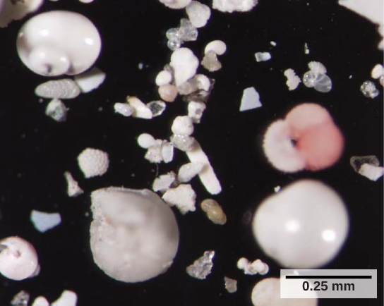
Foraminiferans, or forams, are unicellular heterotrophic protists, ranging from approximately 20 micrometers to several centimeters in length, and occasionally resembling tiny snails ((Figure)). Equally a group, the forams exhibit porous shells, called tests that are built from various organic materials and typically hardened with calcium carbonate. The tests may house photosynthetic algae, which the forams tin harvest for nutrition. Foram pseudopodia extend through the pores and let the forams to move, feed, and gather boosted building materials. Typically, forams are associated with sand or other particles in marine or freshwater habitats. Foraminiferans are also useful as indicators of pollution and changes in global weather patterns.
Foraminiferan Tests. These shells from foraminifera sank to the body of water flooring. (credit: Deep Due east 2001, NOAA/OER)
Radiolarians
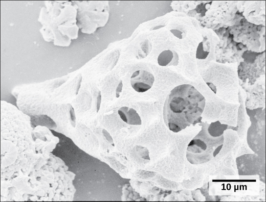
A 2d subtype of Rhizaria, the radiolarians, exhibit intricate exteriors of glassy silica with radial or bilateral symmetry ((Figure)). Needle-like pseudopods supported by microtubules radiate outward from the cell bodies of these protists and office to catch food particles. The shells of dead radiolarians sink to the bounding main flooring, where they may accrue in 100 meter-thick depths. Preserved, sedimented radiolarians are very common in the fossil record.
Radiolarian crush. This fossilized radiolarian shell was imaged using a scanning electron microscope. (credit: modification of work by Hannes Grobe, Alfred Wegener Institute; scale-bar data from Matt Russell)
Cercozoa
The Cercozoa are both morphologically and metabolically diverse, and include both naked and shelled forms. The Chlorarachniophytes ((Figure)) are photosynthetic, having caused chloroplasts by secondary endosymbiosis. The chloroplast contains a remnant of the chlorophyte endosymbiont nucleus, sandwiched between the ii sets of chloroplast membranes. Vampyrellids or "vampire amoebae," every bit their name suggests, obtain their nutrients by thrusting a pseudopod into the interior of other cells and sucking out their contents.
Chromalveolata
Current evidence suggests that species classified as chromalveolates are derived from a common antecedent that engulfed a photosynthetic ruby-red algal cell, which itself had already evolved chloroplasts from an endosymbiotic relationship with a photosynthetic prokaryote. Therefore, the antecedent of chromalveolates is believed to take resulted from a secondary endosymbiotic event. However, some chromalveolates appear to have lost red alga-derived plastid organelles or lack plastid genes altogether. Therefore, this supergroup should be considered a hypothesis-based working group that is bailiwick to change. Chromalveolates include very of import photosynthetic organisms, such equally diatoms, brown algae, and significant disease agents in animals and plants. The chromalveolates can be subdivided into alveolates and stramenopiles.
Alveolates: Dinoflagellates, Apicomplexians, and Ciliates
A large body of information supports that the alveolates are derived from a shared common antecedent. The alveolates are named for the presence of an alveolus, or membrane-enclosed sac, beneath the prison cell membrane. The exact function of the air sac is unknown, but it may exist involved in osmoregulation. The alveolates are further categorized into some of the amend-known protists: the dinoflagellates, the apicomplexans, and the ciliates.
Dinoflagellates exhibit extensive morphological diversity and tin can be photosynthetic, heterotrophic, or mixotrophic. The chloroplast of photosynthetic dinoflagellates was derived by secondary endosymbiosis of a red alga. Many dinoflagellates are encased in interlocking plates of cellulose. Two perpendicular flagella fit into the grooves between the cellulose plates, with one flagellum extending longitudinally and a second encircling the dinoflagellate ((Effigy)). Together, the flagella contribute to the characteristic spinning movement of dinoflagellates. These protists exist in freshwater and marine habitats, and are a component of plankton, the typically microscopic organisms that drift through the water and serve as a crucial nutrient source for larger aquatic organisms.
Dinoflagellates. The dinoflagellates showroom keen diverseness in shape. Many are encased in cellulose armor and have two flagella that fit in grooves between the plates. Motility of these two perpendicular flagella causes a spinning motion.
Dinoflagellates have a nuclear variant chosen a dinokaryon. The chromosomes in the dinokaryon are highly condensed throughout the cell cycle and practice not accept typical histones. Mitosis in dinoflagellates is closed, that is, the spindle separates the chromosomes from exterior of the nucleus without breakdown of the nuclear envelope.
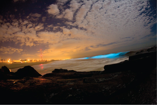
Some dinoflagellates generate lite, chosen bioluminescence, when they are jarred or stressed. Large numbers of marine dinoflagellates (billions or trillions of cells per wave) can emit light and crusade an entire breaking wave to twinkle or take on a brilliant blue color ((Figure)). For approximately xx species of marine dinoflagellates, population explosions (likewise called blooms) during the summertime months tin can tint the ocean with a dingy red colour. This phenomenon is called a carmine tide, and it results from the abundant red pigments present in dinoflagellate plastids. In large quantities, these dinoflagellate species secrete an asphyxiating toxin that tin can kill fish, birds, and marine mammals. Blood-red tides can be massively detrimental to commercial fisheries, and humans who swallow these protists may become poisoned.
Dinoflagellate bioluminescence. Bioluminescence is emitted from dinoflagellates in a breaking wave, as seen from the New Jersey coast. (credit: "catalano82"/Flickr)
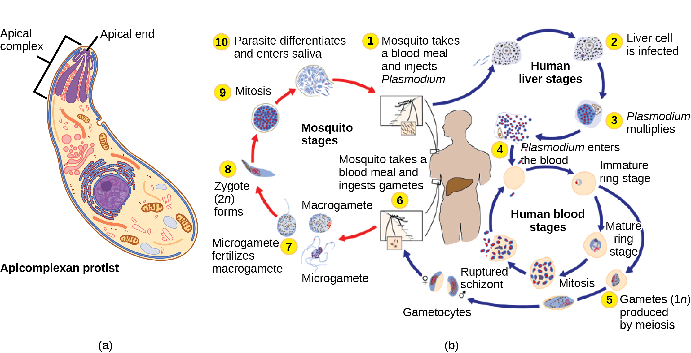
The apicomplexan protists are named for a structure called an apical complex ((Figure)), which appears to be a highly modified secondary chloroplast. The apicoplast genome is like to those of dinoflagellate chloroplasts. The apical circuitous is specialized for entry and infection of host cells. Indeed, all apicomplexans are parasitic. This grouping includes the genus Plasmodium, which causes malaria in humans. Apicomplexan life cycles are complex, involving multiple hosts and stages of sexual and asexual reproduction.
Apicomplexa. (a) Apicomplexans are parasitic protists. They take a characteristic apical complex that enables them to infect host cells. (b) Plasmodium, the causative agent of malaria, has a complex life cycle typical of apicomplexans. (credit b: modification of work by CDC)
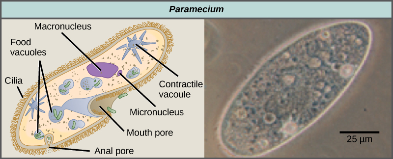
The ciliates, which include Paramecium and Tetrahymena, are a group of protists 10 to 3,000 micrometers in length that are covered in rows, tufts, or spirals of tiny cilia. By beating their cilia synchronously or in waves, ciliates can coordinate directed movements and ingest food particles. Sure ciliates have fused cilia-based structures that role like paddles, funnels, or fins. Ciliates also are surrounded by a pellicle, providing protection without compromising agility. The genus Paramecium includes protists that have organized their cilia into a plate-similar archaic mouth, called an oral groove, which is used to capture and digest bacteria ((Figure)). Nutrient captured in the oral groove enters a food vacuole, where it combines with digestive enzymes. Waste product particles are expelled by an exocytic vesicle that fuses at a specific region on the cell membrane, called the anal pore. In addition to a vacuole-based digestive organisation, Paramecium also uses contractile vacuoles, which are osmoregulatory vesicles that fill with water as information technology enters the jail cell by osmosis and and then contract to clasp h2o from the cell. Ciliates therefore exhibit considerable structural complication without having achieved multicellularity.
Paramecium. Paramecium has a primitive oral cavity (called an oral groove) to ingest food, and an anal pore to eliminate waste matter. Contractile vacuoles allow the organism to excrete excess water. Cilia enable the organism to move. (credit "paramecium micrograph": modification of work by NIH; scale-bar information from Matt Russell)
Link to Learning
Sentinel the video of the contractile vacuole of Paramecium expelling h2o to keep the prison cell osmotically balanced.
Paramecium has two nuclei, a macronucleus and a micronucleus, in each cell. The micronucleus is essential for sexual reproduction, and is in many ways a typical eukaryotic nucleus, except that its genes are not transcribed. The transcribed nucleus is the macronucleus, which directs asexual binary fission and all other biological functions. The macronucleus is a multiploid nucleus constructed from the micronucleus during sexual reproduction. Periodic reconstruction of the macronucleus is necessary because the macronucleus divides amitotically, and thus becomes genetically unbalanced over a menstruum of successive jail cell replications. Paramecium and most other ciliates reproduce sexually by conjugation. This process begins when two dissimilar mating types of Paramecium make physical contact and join with a cytoplasmic bridge ((Figure)). The diploid micronucleus in each cell then undergoes meiosis to produce four haploid micronuclei. Three of these degenerate in each cell, leaving one micronucleus that then undergoes mitosis, generating ii haploid micronuclei. The cells each exchange one of these haploid nuclei and move away from each other. Fusion of the haploid micronuclei generates a completely novel diploid pre-micronucleus in each conjugative prison cell. This pre-micronucleus undergoes iii rounds of mitosis to produce eight copies, and the original macronucleus disintegrates. 4 of the eight pre-micronuclei become full-fledged micronuclei, whereas the other four perform multiple rounds of Dna replication. The copies of the micronuclear chromosomes are severely edited to form hundreds of smaller chromosomes that contain only the protein coding genes. Each of these smaller chromosomes gets new telomeres equally the macronucleus differentiates. Two cycles of jail cell sectionalization then yield four new Paramecia from each original conjugative cell.
Visual Connection
Conjugation in Paramecium. The complex process of sexual reproduction in Paramecium creates viii girl cells from ii original cells. Each jail cell has a macronucleus and a micronucleus. During sexual reproduction, the macronucleus dissolves and is replaced by a micronucleus. (credit "micrograph": modification of work past Ian Sutton; calibration-bar data from Matt Russell)
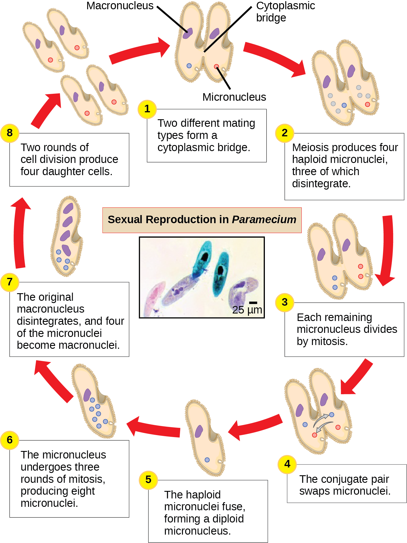
Which of the following statements most Paramecium sexual reproduction is false?
- The macronuclei are derived from micronuclei.
- Both mitosis and meiosis occur during sexual reproduction.
- The conjugate pair swaps macronucleii.
- Each parent produces 4 daughter cells.
<!–<para>C–>
Stramenopiles: Diatoms, Brown Algae, Aureate Algae and Oomycetes
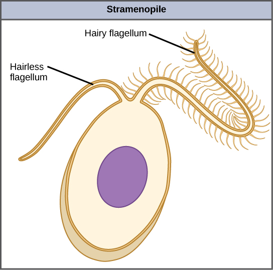
The other subgroup of chromalveolates, the stramenopiles, includes photosynthetic marine algae and heterotrophic protists. The chloroplast of these algae is derived from red alga. The identifying feature of this group is the presence of a textured, or "hairy," flagellum. Many stramenopiles also take an additional flagellum that lacks hair-like projections ((Figure)). Members of this subgroup range in size from single-celled diatoms to the massive and multicellular kelp.
Stramenopile flagella. This stramenopile prison cell has a single hairy flagellum and a secondary smooth flagellum.
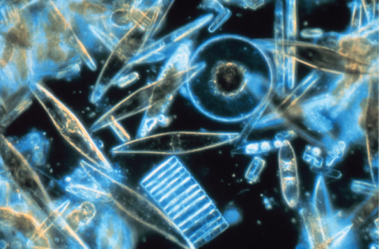
The diatoms are unicellular photosynthetic protists that encase themselves in intricately patterned, glassy jail cell walls composed of silicon dioxide in a matrix of organic particles ((Figure)). These protists are a component of freshwater and marine plankton. Most species of diatoms reproduce asexually, although some instances of sexual reproduction and sporulation also be. Some diatoms exhibit a slit in their silica shell, called a raphe. By expelling a stream of mucopolysaccharides from the raphe, the diatom tin can attach to surfaces or propel itself in 1 direction.
Diatoms. Assorted diatoms, visualized here using low-cal microscopy, live amongst annual sea ice in McMurdo Sound, Antarctica. Diatoms range in size from 2 to 200 µm. (credit: Prof. Gordon T. Taylor, Stony Brook University, NSF, NOAA)
During periods of nutrient availability, diatom populations bloom to numbers greater than can be consumed by aquatic organisms. The excess diatoms die and sink to the sea floor where they are not easily reached past saprobes that feed on dead organisms. Every bit a outcome, the carbon dioxide that the diatoms had consumed and incorporated into their cells during photosynthesis is not returned to the temper. Forth with rhizarians and other shelled protists, diatoms help to maintain a balanced carbon bicycle.
Like diatoms, golden algae are largely unicellular, although some species tin form large colonies. Their feature gold color results from their all-encompassing use of carotenoids, a group of photosynthetic pigments that are more often than not yellow or orangish in colour. Golden algae are institute in both freshwater and marine environments, where they form a major role of the plankton customs.
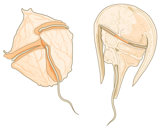
The brown algae are primarily marine, multicellular organisms that are known colloquially every bit seaweeds. Giant kelps are a blazon of brown alga. Some brown algae have evolved specialized tissues that resemble terrestrial plants, with root-like holdfasts, stem-like stipes, and leaf-like blades that are capable of photosynthesis. The stipes of giant kelps are enormous, extending in some cases for lx meters. Like the green algae, brown algae have a multifariousness of life cycles, including alternation of generations. In the brown algae genus Laminaria, haploid spores develop into multicellular gametophytes, which produce haploid gametes that combine to produce diploid organisms that then go multicellular organisms with a dissimilar construction from the haploid grade ((Figure)).
Visual Connection
Alternation of generations in a brown alga. Several species of brown algae, such as the Laminaria shown here, have evolved life cycles in which both the haploid (gametophyte) and diploid (sporophyte) forms are multicellular. The gametophyte is different in structure than the sporophyte. (credit "laminaria photograph": modification of work by Claire Fackler, CINMS, NOAA Photograph Library)
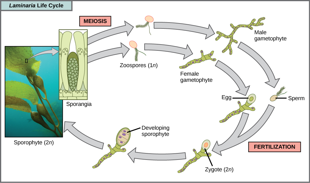
Which of the following statements nearly the Laminaria life cycle is false?
- 1northward zoospores form in the sporangia.
- The sporophyte is the iin plant.
- The gametophyte is diploid.
- Both the gametophyte and sporophyte stages are multicellular.
<!–<para>C–>
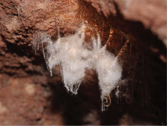
The water molds, oomycetes ("egg fungus"), were so-named based on their fungus-like morphology, but molecular data take shown that the water molds are non closely related to fungi. The oomycetes are characterized by a cellulose-based cell wall and an all-encompassing network of filaments that permit for food uptake. As diploid spores, many oomycetes take two oppositely directed flagella (i hairy and one smooth) for locomotion. The oomycetes are nonphotosynthetic and include many saprobes and parasites. The saprobes appear as white fluffy growths on dead organisms ((Figure)). Most oomycetes are aquatic, simply some parasitize terrestrial plants. One plant pathogen is Phytophthora infestans, the causative agent of late blight of potatoes, such as occurred in the nineteenth century Irish murphy famine.
Oomycetes. A saprobic oomycete engulfs a dead insect. (credit: modification of work by Thomas Bresson)
Excavata
Many of the protist species classified into the supergroup Excavata are asymmetrical, single-celled organisms with a feeding groove "excavated" from one side. This supergroup includes heterotrophic predators, photosynthetic species, and parasites. Its subgroups are the diplomonads, parabasalids, and euglenozoans. The group includes a variety of modified mitochondria, as well as chloroplasts derived from green algae past secondary endosymbiosis. Many of the euglenozoans are free-living, but most diplomonads and parabasalids are symbionts or parasites.
Diplomonads
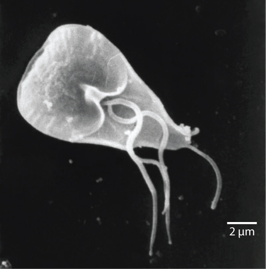
Amongst the Excavata are the diplomonads, which include the intestinal parasite, Giardia lamblia ((Figure)). Until recently, these protists were believed to lack mitochondria. Mitochondrial remnant organelles, chosen mitosomes, take since been identified in diplomonads, but although these mitosomes are essentially nonfunctional as respiratory organelles, they do part in iron and sulfur metabolism. Diplomonads exist in anaerobic environments and utilise alternative pathways, such every bit glycolysis, to generate free energy. Each diplomonad cell has two like, merely non identical haploid nuclei. Diplomonads accept four pairs of locomotor flagella that are adequately deeply rooted in basal bodies that lie betwixt the ii nuclei.
Giardia. The mammalian intestinal parasite Giardia lamblia, visualized here using scanning electron microscopy, is a waterborne protist that causes astringent diarrhea when ingested. (credit: modification of work by Janice Carr, CDC; calibration-bar data from Matt Russell)
Parabasalids
A second Excavata subgroup, the parabasalids, are named for the parabasal appliance, which consists of a Golgi complex associated with cytoskeletal fibers. Other cytoskeletal features include an axostyle, a bundle of fibers that runs the length of the cell and may even extend beyond it. Parabasalids move with flagella and membrane rippling, and these and other cytoskeletal modifications may assist locomotion. Similar the diplomonads, the parabasalids exhibit modified mitochondria. In parabasalids these structures function anaerobically and are called hydrogenosomes because they produce hydrogen gas equally a byproduct.
The parabasalid Trichomonas vaginalis causes trichomoniasis, a sexually transmitted disease in humans, which appears in an estimated 180 one thousand thousand cases worldwide each yr. Whereas men rarely exhibit symptoms during an infection with this protist, infected women may become more susceptible to secondary infection with human being immunodeficiency virus (HIV) and may be more likely to develop cervical cancer. Meaning women infected with T. vaginalis are at an increased chance of serious complications, such as pre-term delivery.
Some of the most complex of the parabasalids are those that colonize the rumen of ruminant animals and the guts of termites. These organisms can digest cellulose, a metabolic talent that is unusual amid eukaryotic cells. They have multiple flagella bundled in circuitous patterns and some additionally recruit spirochetes that adhere to their surface to act every bit accessory locomotor structures.
Link to Learning
Termite gut endosymbionts
Euglenozoans
Euglenozoans includes parasites, heterotrophs, autotrophs, and mixotrophs, ranging in size from 10 to 500 µm. Euglenoids move through their aquatic habitats using two long flagella that guide them toward light sources sensed by a primitive ocular organ chosen an eyespot. The familiar genus, Euglena, encompasses some mixotrophic species that display a photosynthetic adequacy only when low-cal is present. The chloroplast of Euglena descends from a light-green alga by secondary endosymbiosis. In the dark, the chloroplasts of Euglena shrink up and temporarily cease functioning, and the cells instead have up organic nutrients from their environment. Euglena has a tough pellicle equanimous of bands of poly peptide attached to the cytoskeleton. The bands spiral around the cell and give Euglena its exceptional flexibility.
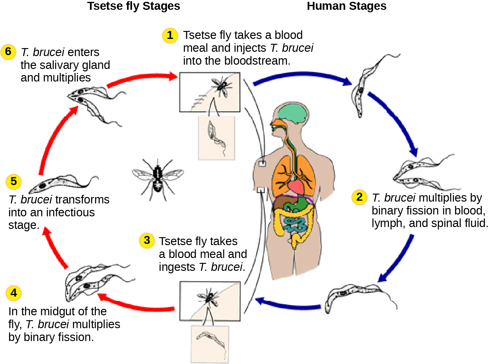
The homo parasite, Trypanosoma brucei, belongs to a different subgroup of Euglenozoa, the kinetoplastids. The kinetoplastid subgroup is named subsequently the kinetoplast, a big modified mitochondrion conveying multiple circular DNAs. This subgroup includes several parasites, collectively called trypanosomes, which cause devastating human diseases and infect an insect species during a portion of their life cycle. T. brucei develops in the gut of the tsetse fly after the fly bites an infected human or other mammalian host. The parasite and then travels to the insect salivary glands to be transmitted to some other human being or other mammal when the infected tsetse fly consumes some other blood meal. T. brucei is common in central Africa and is the causative amanuensis of African sleeping sickness, a disease associated with astringent chronic fatigue, coma, and can be fatal if left untreated.
Sleeping sickness. Trypanosoma brucei, the causative agent of sleeping sickness, spends part of its life cycle in the tsetse fly and part in humans. (credit: modification of work by CDC)
Link to Learning
Watch this video to see T. brucei swimming.
Section Summary
The process of classifying protists into meaningful groups is ongoing, but genetic data in the past twenty years have clarified many relationships that were previously unclear or mistaken. The majority view now is to order all eukaryotes into vi supergroups: Archaeplastida, Amoebozoa, Opisthokonta, Rhizaria, Chromalveolata, and Excavata. The goal of this classification scheme is to create clusters of species that all are derived from a common ancestor. At present, the monophyly of some of the supergroups are better supported by genetic data than others. Although tremendous variation exists within the supergroups, commonalities at the morphological, physiological, and ecological levels can exist identified.
Visual Connection Questions
(Figure) Which of the following statements nearly Paramecium sexual reproduction is false?
- The macronuclei are derived from micronuclei.
- Both mitosis and meiosis occur during sexual reproduction.
- The conjugate pair swaps macronuclei.
- Each parent produces four girl cells.
(Figure) C
(Figure) Which of the post-obit statements about the Laminaria life cycle is false?
- 1n zoospores form in the sporangia.
- The sporophyte is the 2n plant.
- The gametophyte is diploid.
- Both the gametophyte and sporophyte stages are multicellular.
(Figure) C
Review Questions
Which protist grouping exhibits mitochondrial remnants with reduced functionality?
- slime molds
- diatoms
- parabasalids
- dinoflagellates
C
Conjugation between ii Paramecia produces ________ full daughter cells.
- 2
- 4
- 8
- xvi
C
What is the function of the raphe in diatoms?
- locomotion
- defense
- capturing food
- photosynthesis
A
What genus of protists appears to contradict the argument that unicellularity restricts cell size?
- Dictyostelium
- Ulva
- Plasmodium
- Caulerpa
D
A marine biologist analyzing water samples notices a protist with a calcium carbonate trounce that moves past pseudopodia extension. The protist is likely to be closely related to which species?
- Fuligo septica (Dog Vomit slime mold)
- Circogonia icosahedra (Radiolarian)
- Euglena viridis
- Ammonia tepida
D
Disquisitional Thinking Questions
The chlorophyte (green algae) genera Ulva and Caulerpa both have macroscopic leafage-like and stem-similar structures, only but Ulva species are considered truly multicellular. Explicate why.
Unlike Ulva, protists in the genus Caulerpa actually are large, multinucleate, single cells. Because these organisms undergo mitosis without cytokinesis and lack cytoplasmic divisions, they cannot be considered truly multicellular.
Why might a low-cal-sensing eyespot be ineffective for an obligate saprobe? Suggest an alternative organ for a saprobic protist.
By definition, an obligate saprobe lacks the ability to perform photosynthesis, and then it cannot directly obtain nutrition by searching for light. Instead, a chemotactic mechanism that senses the odors released during decay might exist a more constructive sensing organ for a saprobe.
Opisthokonta includes animals and fungi, as well every bit protists. Draw the key feature of this phylum, and an instance of how an organism in each kingdom uses this characteristic.
The central characteristic of Opisthokonts is the flagellum on the posterior stop of cells.
Example organisms:
- Choanoflagellates utilise the flagellum for filter feeding.
- Sponges (animals) use the flagellum for filter feeding.
- Male person gametes (animals) utilise the flagellum for locomotion.
- Fungi spores use the flagellum for locomotion.
Depict ii ways in which paramecium differs from the projected traits of the concluding eukaryotic mutual antecedent.
Possible answers include:
- Two nuclei (a macronucleus and a micronucleus) instead of ane nucleus
- Amitotic partitioning/binary fission during asexual reproduction instead of mitotic jail cell division
- Mitosis of the micronucleus later meiosis instead of direct meiotic production of gametes for sexual reproduction
Glossary
- biological carbon pump
- process by which inorganic carbon is stock-still by photosynthetic species that then die and fall to the sea floor where they cannot be reached by saprobes and their carbon dioxide consumption cannot be returned to the atmosphere
- bioluminescence
- generation and emission of calorie-free by an organism, as in dinoflagellates
- contractile vacuole
- vesicle that fills with water (every bit it enters the cell by osmosis) and so contracts to squeeze h2o from the jail cell; an osmoregulatory vesicle
- cytoplasmic streaming
- movement of cytoplasm into an extended pseudopod such that the unabridged prison cell is transported to the site of the pseudopod
- hydrogenosome
- organelle carried by parabasalids (Excavata) that functions anaerobically and outputs hydrogen gas every bit a byproduct; likely evolved from mitochondria
- kinetoplast
- mass of DNA carried within the unmarried, oversized mitochondrion, characteristic of kinetoplastids (phylum: Euglenozoa)
- mitosome
- nonfunctional organelle carried in the cells of diplomonads (Excavata) that likely evolved from a mitochondrion
- plankton
- diverse grouping of mostly microscopic organisms that drift in marine and freshwater systems and serve as a food source for larger aquatic organisms
- raphe
- slit in the silica shell of diatoms through which the protist secretes a stream of mucopolysaccharides for locomotion and attachment to substrates
- exam
- porous shell of a foram that is built from diverse organic materials and typically hardened with calcium carbonate
Source: https://opentextbc.ca/biology2eopenstax/chapter/groups-of-protists/
Posted by: rosariocreter.blogspot.com
0 Response to "Fungi And Animals Are Both Found In Which Eukaryotic Supergroup?"
Post a Comment